
Central Retinal Vein Occlusion
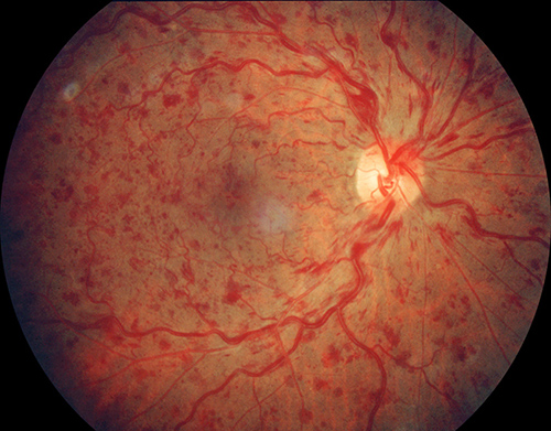
Retinal photograph showing retinal hemorrhages due to a central retinal vein occlusion

Optical coherence tomography (OCT) shows a cross-section of the macula. Leakage of fluid into the retina causes the macula to thicken. The circular areas (arrow) are due to pockets (or cysts) of fluid accumulation within the retina..
Introduction
Please see How the Eye Works for an introduction. Within the retina is a system of blood vessels – arteries, veins, and capillaries. This system of blood vessels nourishes the retina with oxygen. One large artery, the central retinal artery, carries blood to the retina. As the artery enters the eye, many branches spread out throughout the retina. The blood then goes through a fine network of very small vessels called capillaries. After the blood moves through this network of capillaries, it enters branches of veins, called, branch retinal veins. These then join together to form the large vein called the central retinal vein that drains the blood from the eye.
What is a Central Retinal Vein Occlusion? A central retinal vein occlusion or CRVO is a blockage of the main vein that drains blood from the retina. The blockage in this vein causes a back up in blood flow, and increased pressure in the small capillaries.These capillaries then begin to ooze blood and serum. Serum is the clear sticky part of blood. This oozing of fluid into the retina is called retinal edema. Retinal edema and blood in the retina cause the retina to work poorly. The vision becomes blurred and distorted and may have splotches in it. Some blockages are relatively mild and the vision is affected only a little, while others are more severe and the vision is affected much more. Sometimes a CRVO can be so severe, that blood supply to the retina is inadequate and the eye tries to compensate with new blood vessel formation termed neovascularization. Neovascularization can cause bleeding into the eye and an abrupt loss of more vision. Neovascularization can also grow in the front portion of the eye and block the structure that helps to regulate eye pressure. This can cause very high eye pressure and severe pain. This condition is called neovascular glaucoma. A CRVO most often occurs in patients with diabetes, hypertension, and atherosclerosis.
Treatment Occasionally, patients may improve without treatment. However, most people require treatment. Clinical studies have shown that treatment with Lucentis™, Eylea™ and Avastin™ (bevacizumab) are very helpful. These drugs are injected into the eye, and typically given monthly until the retinal swelling resolves, at which point the interval is slowly increased to be sure the swelling doesn’t come back. These drugs are very well tolerated. Although these medications are very effective, most people will suffer at least some loss of vision. Other drugs, Ozurdex™ and triamcinolone can be helpful in many eyes, particularly those that don’t respond to anti-vasogenic drugs These also are injected into the vitreous. Side effects with the steroid drugs can occur, and include cataract formation and glaucoma. These are important to consider with the use of this medication. In contrast, these side-effects are very infrequent with Lucentis, Eylea and Avastin. Laser photocoagulation for CRVO is necessary if the eye develops neovascularization. This is essential to help prevent the complications of neovascular glaucoma.
Copyright 2020 West Coast Retina