Aug, 2023
Presented by Georgia Kamboj, MD, PhD


Presented by Georgia Kamboj, MD, PhD
A 9-year-old boy was referred with a hyperpigmented lesion in the left eye.
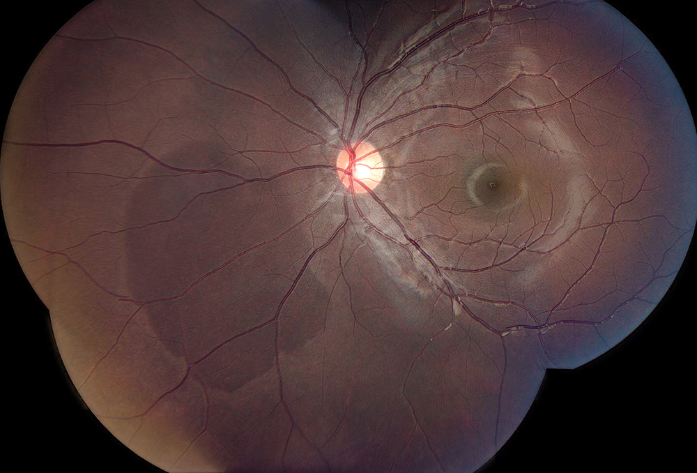
Figure 1: Color photo montage of the left eye. Note the darker area nasal to the nerve
The patient was referred after a routine examination. He denied changes to his vision, flashes/floaters, metamorphopsia, and photophobia. His past ocular history and past medical history were negative. Growth and development were normal for age. Review of systems was negative. He had no relevant family history.
The patient’s best corrected Snellen visual acuity measured 20/20 in each eye. Intraocular pressure was normal in both eyes. Anterior segment exam was unremarkable, without evidence of inflammation. There was no vitritis. Posterior segment exam of the right eye showed no abnormalities. The left fundus was remarkable for a large reddish-brown pigmented patch inferonasal to the disc, approximately 6 mm by 8 mm in diameter (Figure 1). The vasculature appeared normal and there was no subretinal fluid. Macular optical coherence tomography (OCT) of both eyes was unremarkable (Figure 2A-B). OCT taken through the pigmented lesion in the left eye showed hyporeflectivity of the photoreceptor line within the borders of the lesion (Figure 3).

Figure 2A:OCT-SD Scan through the right macula is normal/
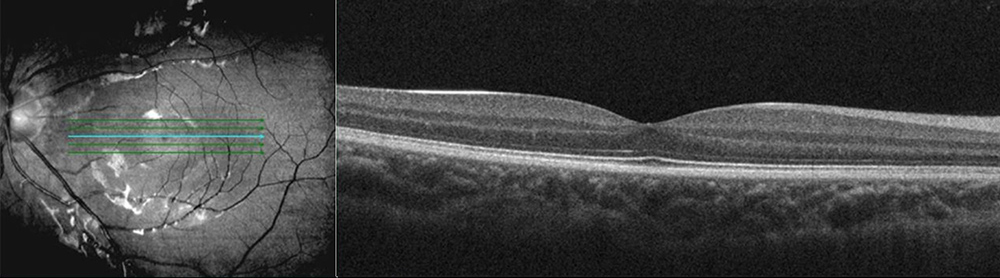
Figure 2B:OCT-SD Scan through the left macula is normal/
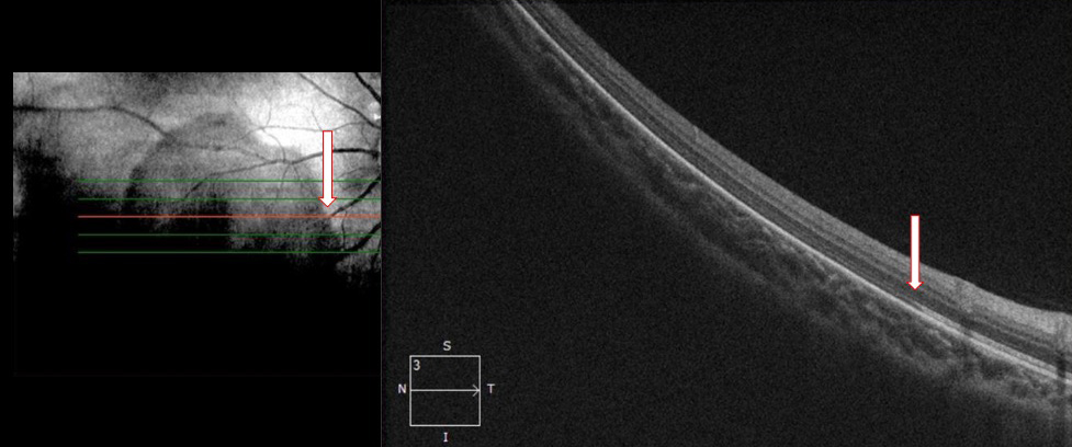
Figure 3:OCT-SD Scan through the lesion. Note the reduced reflectivity in outer retina within the lesion (white arrows)/
Differential Diagnosis
Diagnosis and Patient Course
A diagnosis of dark without pressure was made based on the characteristic OCT findings through the lesion. Observation of the lesions without further workup was recommended.
Discussion
Dark without pressure (DWP) lesions are dark patches of retina found in the central to mid-peripheral fundus. The entity was first described in African patients with hemoglobinopathies 1. DWP are benign lesions that are thought to be acquired during ocular development. They can change shape or completely disappear over time. They do not cause symptoms and are not associated with visual field defects.2 Fawzi et al postulate that the dark lesions represent photopigment with different density or spectral range compared with the rest of the fundus.2 Multimodal imaging shows characteristic findings in DWP lesions. As demonstrated by our case, OCT demonstrates localized hyporeflectivity of the outer segment and ellipsoid zones, with an abrupt change in the reflectivity at the border of the lesion.2,3 Fundus autofluorescence shows hypo-autofluorescence and near infrared reflectance imaging shows hyporeflectivity.2,3