
How the Eye Works
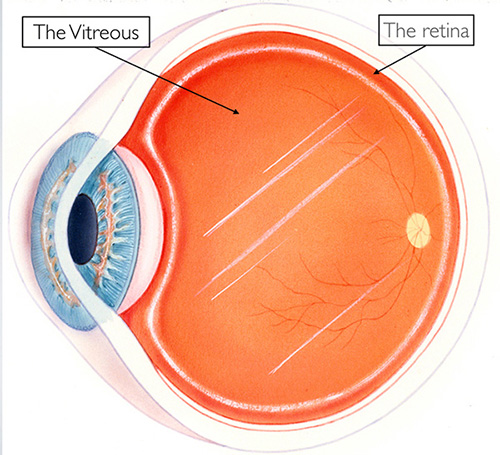
The eye is like a Camera. When you take a picture the lens in the front of the camera allows light through and focuses that light on the film that covers the back inside wall of the camera. When the light hits the film, a picture is taken. The eye works in much the same way. The front parts of the eye (the cornea, pupil, and lens) are clear and allow light to pass through. The light also passes through the large space in the center of the eye called the vitreous cavity. The vitreous cavity is filled with a clear, jelly-like substance called the vitreous or the vitreous gel. The light is focused onto a thin layer of tissue called the retina by the cornea and the lens.
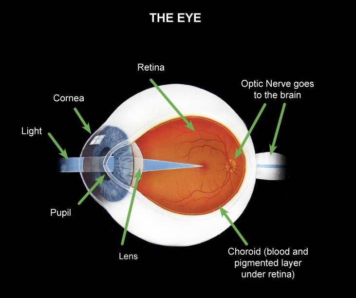
The retina covers the back inside wall of the eye. It is the seeing tissue of the eye. When the focused light hits the retina, a picture is taken. Messages about this picture are sent to the brain through the optic nerve. The brain then interprets these messages and this is how we see.
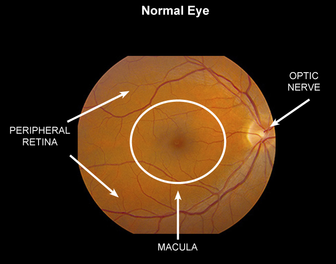
The retina has two parts: the peripheral retina and the macula. If you imagine the retina as a circle with a bull’s-eye at the center, the macula is like the bull’s-eye, it is very small. The large area of the retina that surrounds the macula and makes up 95% of the retina is called the peripheral retina. The peripheral retina gives us vision to the side, called “peripheral” vision. It is this part of the retina that is at work when we see something out of the corner of the eye. Because the peripheral retina is not able to see detail clearly, we cannot use this peripheral vision to read, thread a needle, drive, or even recognize a face. If you see someone off to your side, “out of the corner of your eye”, you may be able to tell who it is because you recognize the person’s general shape, but you won’t be able to see the expression on the person’s face. In order to see fine detail, we must look straight ahead, using the macula, the “bull’s-eye” center of the retina. Even though the macula makes up only a small part of the retina, it is one hundred times more sensitive to detail than the peripheral retina. The macula allows you to see tiny detail, read fine print, recognize faces, thread a needle, read the time, see street signs, and drive a car.

Common Disorders of the Eye
Diagnostic and Retinal Imaging Tests
In order to further evaluate the retina, specialized testing is commonly done including fundus photography, fluorescein angiography and optical coherence tomography (OCT). Other testing, depending on the situation can include indocyanine angiography, fundus autofluorescence and ultrasonography.
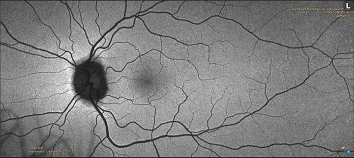
Fundus Photography
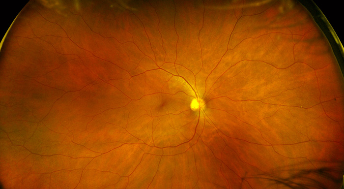
Using specially designed cameras, color photographs of the inside of the eye can be done to assist with diagnosis, and to document findings for comparison with future evaluations. The image on the left reveals more detail, while the image on the right is done with a different camera that can document a larger area of the eye with a single image. These techniques are done depending on the situation.
Optical Coherence Tomography
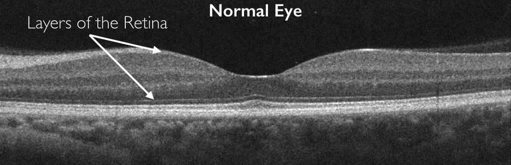
Optical Coherence Tomography (OCT) is probably the most commonly performed diagnostic test of the retina. It allows an extremely detailed evaluation of the layers of the retina. This test is utilized to assist the diagnosis, management and treatment of numerous different eye conditions including diabetic retinopathy, retinal vein occlusions and macular degeneration to name just a few. This test takes only a few minutes and is very well tolerated
Angiography
Fluorescein Angiography-Normal Eye
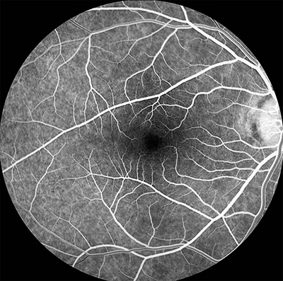
Fluorescein angiography is a very commonly performed test utilizing a special dye that is injected in a vein. This dye travels to the eye and using a special camera, it is possible to evaluate the retinal and choroidal circulation. This dye is unrelated to the common intravenous contrast dyes that are utilized for radiologic studies such as CT scan. Allergies to fluorescein dye are uncommon and the test is very well tolerated. The dye leaves the body through the urine giving it a yellowish-green appearance immediately following the test. This study can be done with a standard camera or a wide-field camera.
Indocyanine Angiography-Normal Eye

Indocyanine angiography is another type of dye study that uses a different type of dye than fluorescein angiography. The properties of this dye show different features in the eye than fluorescein, and is usually used in conjunction with fluorescein angiography in specific situations. It provides complementary information that can not be obtained with fluorescein dye alone. This study can be done with a standard camera or a wide-field camera.
OCT Angiography of Choroidal Neovascularization
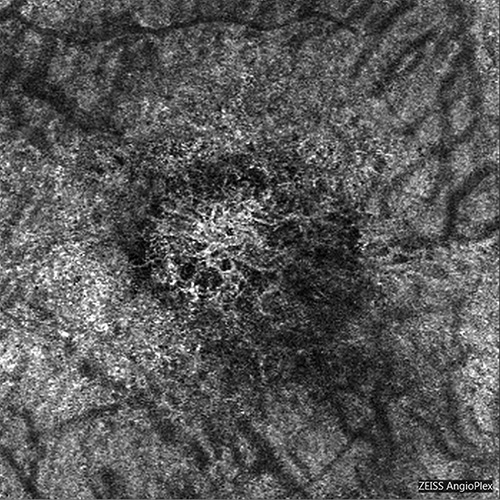
This newly developed technology is a special modification of standard Optical Coherence Tomography that allows visualization of retinal and choroidal vessels in separate layers of the eye. This test takes only a few minutes more in addition to a standard OCT test.
Ultrasonography
Ultrasonogram of Choroidal Tumor

Ultrasonography utilizes sound waves to evaluate tissues. Many people are familiar with this technique to examine pregnant woman. A similar technique can be used to examine the eye as well. This is frequently done to assist in the diagnosis and management of tumors and cancers in the eye. In other situations, the back of the eye can not be adequately examined such as when blood is present. In a situation such as this, ultrasonography is done to determine if the retina is detached.
Fundus Autofluorescence
Wide-Field Autofluorescence Imaging Utilizing a wide-field camera and specialized lighting, pigments in the eye fluoresce when a blue light is shined into the eye. Evaluation of this can be very helpful for diagnosing and following a number of different eye disorders.